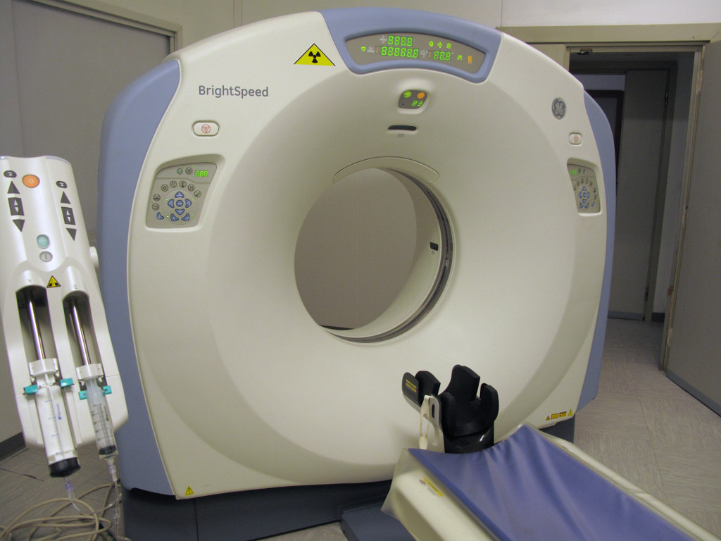
TAC
TC, acronym for computerized tomography, is a diagnostic technique that uses ionizing radiation to get detailed images of specific areas of the body.
The image captured with this method, undergoes a transformation from analog to digital.
During a TC electromagnetic radiation crosses the patient and is captured by detectors (small ionization chambers). In order to obtain detailed information about specific areas of the organism, you need to radiograph the section from multiple angles. The X-ray beam is thus projected following several different trajectories in succession. Such a bundle is much more attenuated as the structures crossed are denser. By constructing a device capable of capturing these differences, you can artificially reconstruct a detailed image of the crossed section.

It is particularly useful in the study of skeletal structures and for analyzing fractures or their outcomes (to evaluate, for example, the position of fracture fragments). It is often used in the oncology field and, thanks to recent developments, is increasingly spreading in the assessment of difficult body areas to investigate such as blood vessels, bronchi, internal heart structures and colon.
No special preparation is required. During TC, body movements should be minimized to avoid blurring images. The patient will however receive appropriate indications from the radiologist. As the examination progresses, the cot advances in small intervals. During a certain or suspect pregnancy it is very important to communicate your condition to your doctor who may eventually decide to postpone the examination or to choose an alternative diagnostic survey. Finally, TC can also be performed in the presence of internal pacemakers or defibrillators.


